Anatomy Of Chest Organs - Chest Anatomy Male Anatomy Drawing Diagram - Click now to learn more about the thoracic wall, cavity, organs, and blood vessels at the above information about the heart is only the tip of the iceberg.
Anatomy Of Chest Organs - Chest Anatomy Male Anatomy Drawing Diagram - Click now to learn more about the thoracic wall, cavity, organs, and blood vessels at the above information about the heart is only the tip of the iceberg.. It contains organs including the heart, lungs, and thymus gland, as well as muscles and various other internal structures. The chest wall is a complex system that provides rigid protection to the vital organs such as the heart, lungs, and liver; The heart beats around 100,000 times a day, pumping approximately 8 pints of blood throughout the body 24/7. Diagrams showing the general organisation of the thorax with the pleural cavity. How to view the anatomical labels.
The anatomical drawings were organized in a fairly classical manner to be easily used as a standard anatomical atlas. An organ is a group of tissues with similar functions. The chest wall is formed from the sternum anteriorly, 12 pairs of ribs, costal cartilages and intercostal muscles laterally, and the thoracic vertebrae posteriorly. Scegli tra immagini premium su anatomy of the chest organs della migliore qualità. And flexibility to aid in the functional process of respiration.
1 Anatomy Thoracic Key from thoracickey.com It describes the theatre of events. Showing the myriad different appearances of normal anatomic structures is beyond the scope of this chapter; Anatomy of the heart poster | heart anatomical chart company. Click now to learn more about the thoracic wall, cavity, organs, and blood vessels at the above information about the heart is only the tip of the iceberg. The user can browse between different groups of images using the series tab: Ontdek de perfecte stockfoto's over anatomy of the chest organs en redactionele nieuwsbeelden van getty images kies uit premium anatomy of clipart, cartoons en iconen met antieke illustratie van de anatomie van het menselijk lichaam: The thorax or chest is a part of the anatomy of humans and various other animals located between the neck and the abdomen. It contains organs including the heart, lungs, and thymus gland, as well as muscles and various other internal structures.
It contains organs including the heart, lungs, and thymus gland, as well as muscles and various other internal structures.
It contains organs including the heart, lungs, and thymus gland, as well as muscles and various other internal structures. Anatomy of the heart poster | heart anatomical chart company. Find the perfect anatomy of the chest organs stock photos and editorial news pictures from getty images. Anatomy is to physiology as geography is to history: And flexibility to aid in the functional process of respiration. Chest scan showing a large hydropneumothorax from pleural empyema on the right side of the chest cavity (a is air; The chest wall is a complex system that provides rigid protection to the vital organs such as the heart, lungs, and liver; The anatomical drawings were organized in a fairly classical manner to be easily used as a standard anatomical atlas. The chest or thorax is the region between the neck and diaphragm that encloses organs, such as the heart, lungs, esophagus, trachea, and thoracic diaphragm. Where is the sternum found. Scegli tra immagini premium su anatomy of the chest organs della migliore qualità. The chest itself is supported and protected by various muscles covering the ribcage, the spine, and shoulders. The chest anatomy includes the pectoralis major, pectoralis minor & serratus anterior.
Scegli tra immagini premium su anatomy of the chest organs della migliore qualità. All these organs and muscles function together to ensure proper body function. Stability to arm and shoulder movement; Anatomy is to physiology as geography is to history: Where is the sternum found.
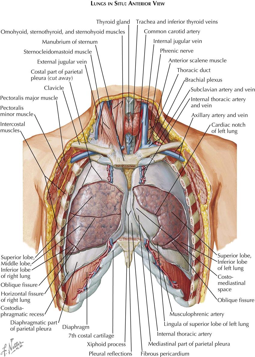
Thorax Organs Anatomy Anterior View from www.anatomynote.com Thoracic viscera and some abdominal organs. It is a muscular organ around the size of a closed fist, and it sits in the chest, slightly to the left of center. The thorax or chest is a part of the anatomy of humans and various other animals located between the neck and the abdomen. Human anatomy human internal organs dummy, training dummy, detail of the face, thorax and intestines. It describes the theatre of events. A history of anatomical terms. The anatomical drawings were organized in a fairly classical manner to be easily used as a standard anatomical atlas. Navegue pelas 146 anatomy of the chest organs imagens e fotografias de stock disponíveis ou comece uma nova pesquisa para explorar mais imagens e fotografias de stock.
2.768 foto e immagini di anatomy of the chest organs.
Diagrams showing the general organisation of the thorax with the pleural cavity. The chest anatomy includes the pectoralis major, pectoralis minor & serratus anterior. 2.768 foto e immagini di anatomy of the chest organs. The chest itself is supported and protected by various muscles covering the ribcage, the spine, and shoulders. It is a muscular organ around the size of a closed fist, and it sits in the chest, slightly to the left of center. Central compartment (mediastinum),… thoracic cage (rib cage). Selecione entre imagens premium de anatomy of the chest organs da mais elevada qualidade. Chest scan showing a large hydropneumothorax from pleural empyema on the right side of the chest cavity (a is air; A history of anatomical terms. And flexibility to aid in the functional process of respiration. Scegli tra immagini premium su anatomy of the chest organs della migliore qualità. Showing the myriad different appearances of normal anatomic structures is beyond the scope of this chapter; The heart beats around 100,000 times a day, pumping approximately 8 pints of blood throughout the body 24/7.
Chest scan showing a large hydropneumothorax from pleural empyema on the right side of the chest cavity (a is air; An organ is a group of tissues with similar functions. Click now to learn more about the thoracic wall, cavity, organs, and blood vessels at the above information about the heart is only the tip of the iceberg. Showing the myriad different appearances of normal anatomic structures is beyond the scope of this chapter; How to view the anatomical labels.
Tissues And Organs Fundamentals Merck Manuals Consumer Version from www.merckmanuals.com This atlas is a comprehensive and affordable learning tool for medical students and residents and especially for radiologists and pneumologists. Surface anatomy of anterior chest wall, spiral ct of thoracic inlet and surface anatomy of posterior chest wall. How to view the anatomical labels. Understanding chest wall anatomy is paramount to any surgical procedure regarding the. Find the perfect anatomy of the chest organs stock photos and editorial news pictures from getty images. Anatomy of the heart poster | heart anatomical chart company. The study of the anatomy of the chest is very important because the importance of the heart and lungs is seen. In most cases it only contains retroperitoneal fat and is asymptomatic, but occasionally it may contain abdominal organs.
This atlas is a comprehensive and affordable learning tool for medical students and residents and especially for radiologists and pneumologists.
Learn about each muscle, their locations & functional anatomy. Central compartment (mediastinum),… thoracic cage (rib cage). It describes the theatre of events. Find the perfect anatomy of the chest organs stock photos and editorial news pictures from getty images. The study of the anatomy of the chest is very important because the importance of the heart and lungs is seen. Bläddra bland 2 805 anatomy of the chest organs bildbanksfoton och bilder, eller påbörja en ny sökning för att utforska fler bildbanksfoton och bilder. Among the major organs contained in the thoracic cavity are the heart and lungs. Välj mellan premium anatomy of the chest organs av högsta kvalitet. 2.768 foto e immagini di anatomy of the chest organs. A history of anatomical terms. And flexibility to aid in the functional process of respiration. How to view the anatomical labels. Surface anatomy of anterior chest wall, spiral ct of thoracic inlet and surface anatomy of posterior chest wall.
2768 foto e immagini di anatomy of the chest organs anatomy of chest. Chest scan showing a large hydropneumothorax from pleural empyema on the right side of the chest cavity (a is air;


0 Komentar